


The Principle of a Wound Healing Assay and Migration Assay
Applications of Wound Healing and Migration Assays
Cell migration occurs during many important physiological processes. For example, controlled cell migration allows for embryonic development, tissue injury, and wound healing. In contrast, cell migration is dysregulated in many pathological situations, such as cancer metastasis and inflammation. Not surprisingly, the principles behind cell migration have been studied in many contexts.
Wound healing assays and migration assays are widely used approaches for the analysis of cell migration under different conditions. They give insight into the following relevant scientific topics:
- Conditional Migration and Wound Healing Studies: How do the cells migrate under different conditions (e.g., after being treated with a specific compound/enhancer/inhibitor, after gene silencing using siRNA or CRISPR/Cas9, after changing substrate stiffness, or after changing matrix composition)?
- High Throughput Drug Screening: Which drug alters the migration rate of a specific cell type (e.g., cancer cells)?
- Cell Interaction Studies: Do the cells remain in contact or do they migrate singularly? How do the cells interact with each other during migration? What is the cell-autonomous migration ability?
- 2D Invasion Assays: How do two different cell types interact with each other (e.g., tumor cells and fibroblasts)?
In contrast to general wound healing and migration assays, directed migration assays, such as chemotaxis assays, measure a gradient-dependent cell movement in a 2D or 3D environment. Please find more information on chemotaxis assays here.

Endothelial cells cultured in a µ-Dish 35 mm, high with a Culture-Insert 2 Well. Image by Derek Sung, University of Pennsylvania School of Medicine, USA.

Two different cell types in a 2D invasion assay. Data provided by C. Matern and K. D. Nnetu, University Leipzig, Germany.
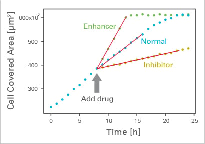
Example analysis of the influence of inhibitors or enhancers on wound healing.
The most characteristic readout of a wound healing assay / migration assay is the change of the cell-covered area (gap closure) over time. Conducting a wound healing and migration assay is an easy procedure:
- Create a physical gap within a cell monolayer.
- Monitor the process of cell migration into the gap with live cell imaging or by taking photos at different time points.
- Analyze the gap closure rate, which is a typical experimental readout, manually or by using automated software.
Despite the apparent simplicity of a wound healing assay, many factors can influence the experimental outcome and they have to be controlled. In the following sections, you will learn about the principle and the setup of a wound healing assay. In addition, we will discuss the parameters that need to be standardized in order to achieve reproducible, robust results.

Live cell imaging using the ibidi Stage Top Incubation System shows the gap closure of MCF7 cells in a wound healing and migration assay. Phase contrast; 10x objective lens.
J.E.N. Jonkman, et al. An introduction to the wound healing assay using live-cell microscopy. Cell Adh Migr, 2014, 10.4161/cam.36224
read abstract
W.J. Ashby, A. Zijlstra. Established and novel methods of interrogating two-dimensional cell migration. Integr Biol 2012, 10.1039/c2ib20154b
read abstract
Read on and learn more about the Experimental Workflow of a wound healing assay or different Methods of Creating the Gap.